
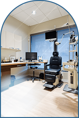
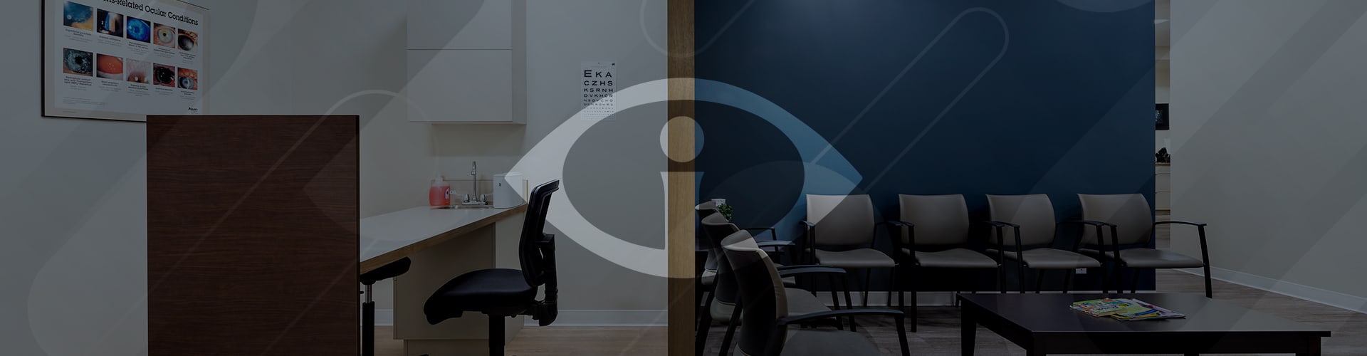
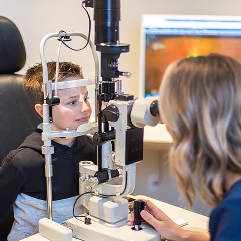
Our Technology
At iSight Optometry, we utilize modern technology to help diagnose and manage various eye diseases, such as glaucoma, age-related macular degeneration, and keratoconus. These tools help us to create treatment plans for our patients and offer comprehensive vision care.
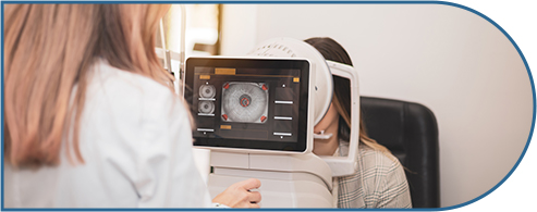
Autorefraction
Autorefraction can provide a quick and accurate measurement of how light refracts in your eye, helping to determine your glasses or contact lens prescription.
Noncontact Tono-Pachymetry (NCT Pachymetry)
NCT Pachymetry is a noninvasive technology that measures the eye pressure as well as thickness of the cornea. It can help detect glaucoma and other eye diseases.
Topography
Corneal topography creates a map of your cornea’s surface, which helps diagnose and manage eye conditions like keratoconus and fitting specialty contact lenses.
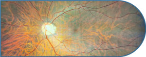
Optos Widefield Imaging
Optomap Ultra-Widefield digital imaging is an exciting technology that allows you and your optometrist to see a 200-degree picture or image of the inside of your eye. It allows our optometrists to show you areas of concern that might need monitoring or treatment. It also allows our optometrists to detect even the earliest signs of diseases that appear on your retina. We Include Optomap imaging as part of every comprehensive eye exam.
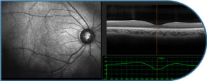
Optical Coherence Tomography (OCT)
Optical coherence tomography technology creates cross-sectional images of the retina, optic nerve, and macula, which can help to diagnose age-related macular degeneration and glaucoma.
Visual Field Testing
Visual Field Testing assesses both peripheral and central vision, helping to detect neurological disorders that can affect vision.
MacuMira
MacuMira is a clinically proven and Health Canada-approved treatment option that can provide hope for those seeking to slow the progression of dry AMD and potentially improve their vision. MacuMira is a safe and noninvasive treatment that uses microcurrent stimulation to target the retinal cells in your eye. This gentle stimulation is designed to help:
• Enhance the function of the retinal pigment epithelium (RPE), a layer crucial for removing waste products from the macula
• Improve blood flow and nourish the macula, the central area responsible for sharp, central vision
• Stimulate the production of essential cellular components that promote retinal health.
We Make Our Technology Work for You
Managing your eye health requires a collaborative effort. We leverage our technology to empower and educate our patients during their appointments.
At iSight Optometry, we want our patients to be informed and involved in their eye care journey. Our tools help us to promote understanding and engagement in our patients and their eye health.
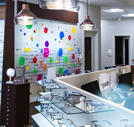
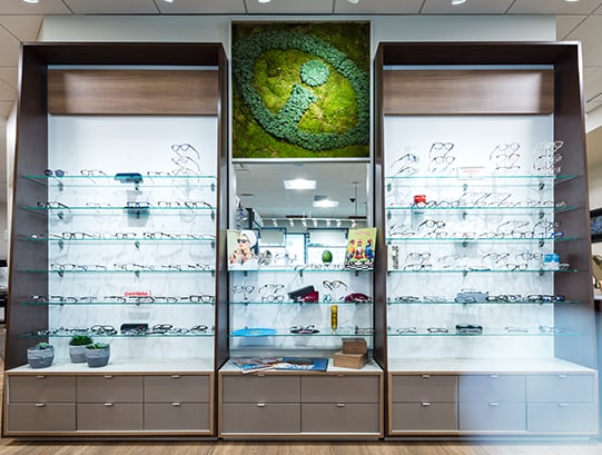
Providing the Correct Fit for Your Glasses & Lenses
Optikam is a modern optical technology used at iSight Optometry to help fit eyeglasses to your face. The device captures a number of measurements–like your pupillary distance–and creates a 3D image of your face.
Optikam can enhance comfort by providing accurately aligned lenses and creating a more efficient fitting experience. This technology also helps tailor eyewear to your unique facial features.
Our in-office edger allows us to craft eyeglasses on-site, which helps us custom-fit lenses for our patients.
Book an Eye Exam with Us Today
Our technology can enhance your eye care experience. Contact us to schedule your appointment today.
Book Appointment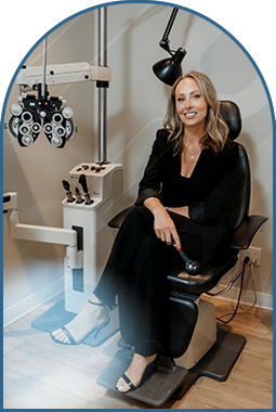
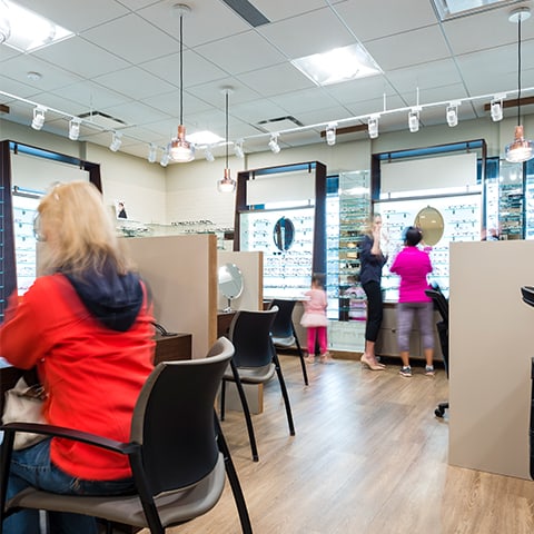
Come Visit Us
Visit Us
We are on Harvey Avenue, and there is plenty of parking outside the clinic. Our location is wheelchair accessible! We look forward to welcoming you and your family.
Where to park?
Our Address
- 4 – 2070 Harvey Avenue
- Kelowna, BC V1Y 8P8
Contact Information
- Phone: 250-860-2020
- Fax: 250-762-9411
- Email: [email protected]
Our Hours
- Monday: 8:00 AM – 5:00 PM
- Tuesday: 8:00 AM – 5:00 PM
- Wednesday: 8:00 AM – 5:00 PM
- Thursday: 8:00 AM – 5:00 PM
- Friday: 8:00 AM – 2:30 PM
- Saturday: Closed
- Sunday: Closed
Our Google Reviews
Our Brands
-
iSight Optometry
- 4 - 2070 Harvey Avenue
- Kelowna, BC V1Y 8P8
-
P: 250-860-2020
[email protected]